- 移动端


北京隆福佳生物科技有限公司代理商
8 年
手机商铺
商家活跃:
产品热度:
- NaN
- 0.8999999999999999
- 0.8999999999999999
- 2.9
- 2.9

整合 PE 系列产品的低氧/厌氧环境模拟系统
询价
代理商
北京隆福佳生物科技有限公司
入驻年限:8 年
- 联系人:
纪小姐
- 所在地区:
北京 石景山区
- 业务范围:
技术服务、实验室仪器 / 设备、细胞库 / 细胞培养、试剂、ELISA 试剂盒、抗体、耗材、论文服务、医疗器械
- 经营模式:
经销商 代理商
推荐产品


公司新闻/正文
低氧/厌氧产品案例——低氧与视网膜病变
645 人阅读发布时间:2022-05-26 10:55
文章题目:Long non-coding RNA histone deacetylase 4 antisense RNA 1 (HDAC4-AS1) inhibits HDAC4 expression in human ARPE-19 cells with hypoxic stress
长非编码 RNA 组蛋白去乙酰化酶 4 反义 RNA 1 (HDAC4-AS1) 抑制低氧胁迫下人 ARPE-19 细胞 HDAC4 的表达
文章出处:Bioengineered, 2021, 12: 2228-2237; 中国山东省淄博市中心医院眼科
工作站使用情况:InVivo2 400
使用气体 浓度:低氧(<0.1% O2)
摘要:年龄相关性黄斑变性 (AMD) 是由脉络膜新生血管 (CNV) 介导的瘢痕形成和视力丧失引起的。据报道,视网膜色素上皮 (RPE) 细胞中持续的视网膜缺氧导致了 CNV。然而,视网膜色素上皮细胞缺氧反应的潜在基因调控网络尚未完全了解。在本研究中,人 ARPE-19 视网膜色素上皮细胞在缺氧条件下培养 24 小时,然后在常氧条件下复氧。然后通过高通量测序研究转录组。我们观察到长非编码 RNA (lncRNA) 组蛋白去乙酰化酶 4 反义 RNA 1 (HDAC4-AS1) 在低氧条件下比正常对照组增加,在添加复氧后减少,而 HDAC4 表达的变化在低氧条件下比正常对照组减少,在 ARPE-19 细胞中添加复氧后上调。此外,HDAC4-AS1 敲除只能在缺氧条件下抑制 HDAC4 的转录活性,荧光原位杂交和 pull down 实验表明 HDAC4-AS1 转录物能与 HDAC4 启动子结合并促进 HIF-1α 的募集。最后,我们还确定了 HDAC4-AS1 与 HIF-1α 和 HDAC4 启动子相互作用的特定区域。综上所述,这些结果表明 HDAC4- AS1 可以通过调节缺氧应激的人 ARPE-19 细胞中 HIF-1α 来抑制 HDAC4 的表达。
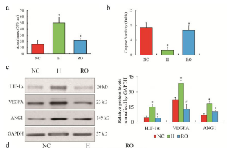
Figure 1. The RNA profiles of ARPE-19 cells treated by hypoxia. The phenotype of cell growth (a) and apoptosis (b) of cultured ARPE19 cells under hypoxic and re-oxygenated conditions. The protein expression of HIF-1α, VEGFA and ANG1 in ARPE-19 cells (c).
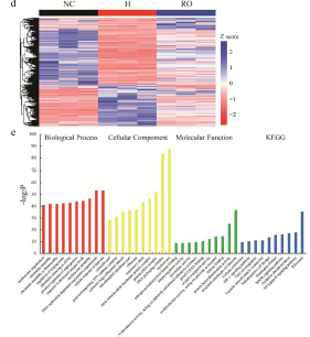
Figure 1 |continue. Heatmap of differential expressed genes of ARPE-19 cells (d). Color bars above the heatmap represent sample groups: red is for upregulated genes and blue is for down-regulated genes. Gene ontology analysis including biological process, cellular component and molecular function and KEGG analysis (e) of the top -10 function enrichments or pathways associated with these differential expressed genes. NC means normoxic condition, H means hypoxic condition and RO means re-oxygenation. The comparison of H to NC (『*』) and RO to H (『#』) with the statistical significance that p value is less than 0.05. Data are presented as mean ± standard error of the mean of three individual experiments.
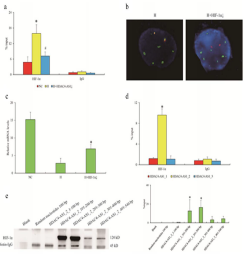
Figure 3.The interaction between HDAC4-AS1 and HIF-1α in ARPE- 19 cells. Enrichment of HIF-1α on shared promoter of HDAC4/HDAC4-AS1 shown by ChIP-qPCR assay (a). The statistical significance by the comparison between H and NC (『*』) as well as
between H+ HDAC4-AS1 knockdown and H (『#』), with p value less than 0.05. The interaction between HDAC4-AS1 and promoter region affected by HIF-1α knockdown shown by FISH assay (b). The HDAC4 transcription affected by HIF-1α knockdown (c). 『*』 represents the statistical significance by the comparison between H + HIF-1α knockdown and H, with p value less than 0.05. The interaction between HIF-1α and different variants of HDAC4-AS1 (d). 『*』 represents the statistical significance comparing HIF-1α with IgG in HDAC4-AS1_2 group. The detailed binding domain of HDAC4-AS1_2 interacted with HIF-1α shown by pull down and western blot assays (e).
ARPE-19 细胞在低氧 (< 0.1% O2,5% CO2 和 95% N2) 或常氧 (21% 氧) 培养 24 小时,然后复氧 48 h;
缺氧 (H) 可增强 ARPE 细胞的增殖能力,抑制凋亡,而复氧则可削弱其增殖能力,促进凋亡 (图 1a, b);HIF-1α、VEGFA 和 ANG1 在低氧时高表达,复氧表达下调 (图 1c);接下来,对 NC、H 和 RO 组的 RNA 图谱进行了表征,发现 HDAC4-AS1 可能是响应 ARPE-19 细胞氧浓度的新候选者之一(图 1d,e);
在低氧条件下,HDAC4- as1 基因的敲除会削弱其与 HDAC4/HDAC4- as1 启动子的相互作用 (图 3a);HIF-1α 的下调可能会影响 HDAC4-AS1 与启动子区的相互作用 (图 3b),减弱 HDAC4 转录 (图 3c),表明 HIF-1α 可能在 HDAC4-AS1 调控的 HDAC4 的抑制调控中发挥关键作用。
本研究揭示在缺氧的 ARPE-19 细胞中,HDAC4- as1 通过招募 HIF-1α 来抑制 HDAC4 转录活性。
长非编码 RNA 组蛋白去乙酰化酶 4 反义 RNA 1 (HDAC4-AS1) 抑制低氧胁迫下人 ARPE-19 细胞 HDAC4 的表达
文章出处:Bioengineered, 2021, 12: 2228-2237; 中国山东省淄博市中心医院眼科
工作站使用情况:InVivo2 400
使用气体 浓度:低氧(<0.1% O2)
摘要:年龄相关性黄斑变性 (AMD) 是由脉络膜新生血管 (CNV) 介导的瘢痕形成和视力丧失引起的。据报道,视网膜色素上皮 (RPE) 细胞中持续的视网膜缺氧导致了 CNV。然而,视网膜色素上皮细胞缺氧反应的潜在基因调控网络尚未完全了解。在本研究中,人 ARPE-19 视网膜色素上皮细胞在缺氧条件下培养 24 小时,然后在常氧条件下复氧。然后通过高通量测序研究转录组。我们观察到长非编码 RNA (lncRNA) 组蛋白去乙酰化酶 4 反义 RNA 1 (HDAC4-AS1) 在低氧条件下比正常对照组增加,在添加复氧后减少,而 HDAC4 表达的变化在低氧条件下比正常对照组减少,在 ARPE-19 细胞中添加复氧后上调。此外,HDAC4-AS1 敲除只能在缺氧条件下抑制 HDAC4 的转录活性,荧光原位杂交和 pull down 实验表明 HDAC4-AS1 转录物能与 HDAC4 启动子结合并促进 HIF-1α 的募集。最后,我们还确定了 HDAC4-AS1 与 HIF-1α 和 HDAC4 启动子相互作用的特定区域。综上所述,这些结果表明 HDAC4- AS1 可以通过调节缺氧应激的人 ARPE-19 细胞中 HIF-1α 来抑制 HDAC4 的表达。
Figure 1. The RNA profiles of ARPE-19 cells treated by hypoxia. The phenotype of cell growth (a) and apoptosis (b) of cultured ARPE19 cells under hypoxic and re-oxygenated conditions. The protein expression of HIF-1α, VEGFA and ANG1 in ARPE-19 cells (c).
Figure 1 |continue. Heatmap of differential expressed genes of ARPE-19 cells (d). Color bars above the heatmap represent sample groups: red is for upregulated genes and blue is for down-regulated genes. Gene ontology analysis including biological process, cellular component and molecular function and KEGG analysis (e) of the top -10 function enrichments or pathways associated with these differential expressed genes. NC means normoxic condition, H means hypoxic condition and RO means re-oxygenation. The comparison of H to NC (『*』) and RO to H (『#』) with the statistical significance that p value is less than 0.05. Data are presented as mean ± standard error of the mean of three individual experiments.
Figure 3.The interaction between HDAC4-AS1 and HIF-1α in ARPE- 19 cells. Enrichment of HIF-1α on shared promoter of HDAC4/HDAC4-AS1 shown by ChIP-qPCR assay (a). The statistical significance by the comparison between H and NC (『*』) as well as
between H+ HDAC4-AS1 knockdown and H (『#』), with p value less than 0.05. The interaction between HDAC4-AS1 and promoter region affected by HIF-1α knockdown shown by FISH assay (b). The HDAC4 transcription affected by HIF-1α knockdown (c). 『*』 represents the statistical significance by the comparison between H + HIF-1α knockdown and H, with p value less than 0.05. The interaction between HIF-1α and different variants of HDAC4-AS1 (d). 『*』 represents the statistical significance comparing HIF-1α with IgG in HDAC4-AS1_2 group. The detailed binding domain of HDAC4-AS1_2 interacted with HIF-1α shown by pull down and western blot assays (e).
ARPE-19 细胞在低氧 (< 0.1% O2,5% CO2 和 95% N2) 或常氧 (21% 氧) 培养 24 小时,然后复氧 48 h;
缺氧 (H) 可增强 ARPE 细胞的增殖能力,抑制凋亡,而复氧则可削弱其增殖能力,促进凋亡 (图 1a, b);HIF-1α、VEGFA 和 ANG1 在低氧时高表达,复氧表达下调 (图 1c);接下来,对 NC、H 和 RO 组的 RNA 图谱进行了表征,发现 HDAC4-AS1 可能是响应 ARPE-19 细胞氧浓度的新候选者之一(图 1d,e);
在低氧条件下,HDAC4- as1 基因的敲除会削弱其与 HDAC4/HDAC4- as1 启动子的相互作用 (图 3a);HIF-1α 的下调可能会影响 HDAC4-AS1 与启动子区的相互作用 (图 3b),减弱 HDAC4 转录 (图 3c),表明 HIF-1α 可能在 HDAC4-AS1 调控的 HDAC4 的抑制调控中发挥关键作用。
本研究揭示在缺氧的 ARPE-19 细胞中,HDAC4- as1 通过招募 HIF-1α 来抑制 HDAC4 转录活性。