- 移动端


北京隆福佳生物科技有限公司代理商
8 年
手机商铺
- NaN
- 0
- 1
- 0
- 3

北京隆福佳生物科技有限公司
入驻年限:8 年
- 联系人:
纪小姐
- 所在地区:
北京 石景山区
- 业务范围:
技术服务、实验室仪器 / 设备、细胞库 / 细胞培养、试剂、ELISA 试剂盒、抗体、耗材、论文服务、医疗器械
- 经营模式:
经销商 代理商
推荐产品


公司新闻/正文
低氧/厌氧产品案例——低氧与脑血管内皮细胞研究
327 人阅读发布时间:2021-12-15 11:04
文章题目:Nrf2-regulated redox signaling in brain endothelial cells adapted to physiological oxygen levels: Consequences for sulforaphane mediated protection against hypoxia reoxygenation
适应生理氧水平的脑内皮细胞中Nrf2 调节的氧化还原信号:萝卜硫素介导的缺氧-复氧保护
文章出处:RedoxBiology37(2020)101708.英国牛津大学纳菲尔德医学系目标发现研究所
工作站使用情况:SCI-tive Dual
使用气体浓度:1Kpa,5Kpa,18Kpa
摘要:缺血性中风与再灌注过程中活性氧生成激增有关。静脉溶栓和血管内取栓的狭窄治疗窗口限制了患者的治疗选择。因此,了解调节神经血管氧化还原防御的机制是改善临床问题的关键。我们前期在啮齿类动物缺血脑卒中模型中的研究证实,萝卜硫素(SFN)预处理激活Nrf2 防御酶,对神经血管和神经功能缺损具有保护作用。我们进一步研究了SFN 介导的小鼠大脑微血管内皮细胞(bEnd.3)对高氧(18 kPa)和常氧(5 kPa) O2 水平的长期(5 天)保护作用。利用O2 敏感的磷光纳米粒子探针,我们测量了在5kpa O2 条件下培养的bEnd 3 细胞的细胞内氧水平为3.4±0.1 kPa。2.5 μM SFN 对HO-1 和GCLM 的诱导在5kpa O2 的细胞中显著减弱,尽管Nrf2 在细胞核中积累。大脑微血管内皮细胞模拟缺血性中风模型。3 个细胞适应18 或5kpa 氧气,并接受缺氧(1kpa 氧气,1 h)和复氧。在适应18kpa O2 的细胞中,PEG-SOD 能消除复氧诱导的自由基产生,而2.5 μM SFN 预处理能明显减弱。Nrf2 转录沉默抑制了HO-1 和NQO1 的诱导,导致复氧诱导的自由基产生显著增加。值得注意的是,使用发光探针L-012 和荧光探针MitoSOX™Red 和FeRhoNox™-1 检测,在5kpa 氧气条件下培养的细胞中,复氧诱导的氧化应激减少,表明适应生理常氧的脑微血管细胞氧化还原表型改变。由于氧化还原和其他细胞内信号通路受到O2 的严重影响,针对Keap1-Nrf2 de通路治疗脑卒中、冠状动脉和肾脏疾病缺血-再灌注损伤的抗氧化疗法的开发,需要在明确的O2水平下进行体外研究。
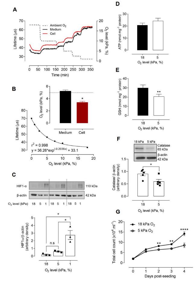
Fig. 2.Adaptation to 5 kPa O2 alters the redox phenotype of bEnd.3 cells in the absence of HIF-1α stabilization bEnd.3 cells adapted to 18 kPa O2 were loaded with MitoXpress®-INTRA for 16 h, transferred rapidly to an O2-regulated plate reader and exposed to stepwise reductions in O2 (dotted line, right axis). (A) Phosphores cence lifetime measurements (see Methods) in cells and dissolved O2 in medium. (B) Averaged phosphorescence lifetime versus ambient O2 levels in the plate reader were fit by exponential analysis. Inset: Interpolated O2 content in bEnd.3 cell cytosol and me dium under 5 kPa O2 (dashed line).(C) Im munoblots of HIF-1α expression relative to β- actin and densitometric analysis of 3 cul tures (separated by dashed lines). (D–E) Intracellular ATP and GSH levels in cells adapted for 5 d to 18 or 5 kPa O2. (F) Immunoblot and densitometric analysis of catalase expression relative to β-actin under 18 or 5 kPa O2. (G) Differential rate of bEnd.3 cell proliferation under 18 or 5 kPa O2. Data denote mean ±S.E.M., n =3–5 independent bEnd.3 cell cultures, *P <0.05, **P <0.01, ****P <0.0001, n.s. nonsignificant.
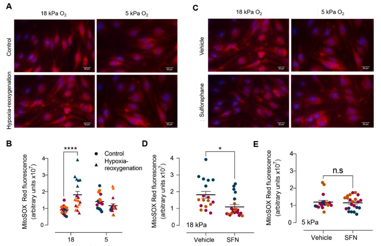
Fig. 6.Effects of sulforaphane pretreatment on reoxygenation induced mitochondrial reactive oxygen species generation in bEnd.3 cells adapted to 18 kPa or 5 kPa O2 bEnd.3 cells seeded in Ibidi μ-Slide 8-well chambers were cultured under 18 or 5 kPa O2 for 5 d. Cells were subjected to hypoxia (1 kPa O2, 1 h) and loaded with MitoSOX™ Red for 5 min before the start of 30 min reoxygenation under 18 or 5 kPa O2, respectively. Control cells were loaded with MitoSOX™ Red during the last 30 min of an experiment. Cells were fixed with 4% paraformaldehyde and images acquired using a Nikon Diaphot microscope with a 40×objective. (A) Repre sentative images of MitoSOX fluorescence and DAPI stained nuclei after 30 min reoxygenation and (B) quantification of MitoSOX fluorescence. (C) bEnd.3 cells were pre-treated with vehicle (0.01% DMSO) or SFN (2.5 μM) for 24 h before exposure to hypoxia (1 h) and reoxygenation under 18 or 5 kPa O2, respectively. Repre sentative images of MitoSOX fluorescence and DAPI stained nuclei after 30 min reoxygenation and (D–E) quantitation of MitoSOX fluorescence. Each symbol in panels B,
D and E represents the mean fluorescence from at least 10 cells in a field of view, with each color denoting a different bEnd.3 experiment with at least 6 different fields of view. Data denote mean ±S.E.M., n =18–20 fields of view in each of 3 independent bEnd.3 cell cultures, two-way ANOVA followed by Bonferroni post-hoc analysisRepresentative blots are presented.
在5kpa 氧气条件下培养的细胞中,细胞内的氧气水平为3.4±0.1 kPa(见图2B),与清醒小鼠[41]皮层中的水平相似(图2B);3 个细胞适应于18kpa、5 kPa 或1kpa 的氧气,HIF-1α仅在1kpa 缺氧条件下检测到稳定表达(图2C),在高氧(18kpa O2)条件下标准细胞培养过程中增强的氧化应激在适应生理常氧5kpa O2 的细胞中减弱(图2D-F)。表明5kpa O2 适应改变了大脑微血管内皮细胞的氧化还原表型;缺氧复氧诱导MitoSOX 红荧光增加,2.5 μM SFN 预处理12 h 可显著降低细胞复氧诱导的MitoSOX红荧光增加(图6),表明萝卜硫素预处理减少大脑微血管内皮细胞复氧诱导的线粒体活性氧的生成。