- 移动端


北京隆福佳生物科技有限公司代理商
8 年
手机商铺
商家活跃:
产品热度:
- NaN
- 0.8999999999999999
- 0.8999999999999999
- 2.9
- 2.9

整合 PE 系列产品的低氧/厌氧环境模拟系统
询价
代理商
北京隆福佳生物科技有限公司
入驻年限:8 年
- 联系人:
纪小姐
- 所在地区:
北京 石景山区
- 业务范围:
技术服务、实验室仪器 / 设备、细胞库 / 细胞培养、试剂、ELISA 试剂盒、抗体、耗材、论文服务、医疗器械
- 经营模式:
经销商 代理商
推荐产品


公司新闻/正文
低氧/厌氧产品案例——低氧与血管新生研究
526 人阅读发布时间:2022-05-18 16:39
文章题目:LTBP4 affects renal fibrosis by influencing angiogenesis and altering mitochondrial structure
LTBP4 通过影响血管新生和改变线粒体结构影响肾纤维化
文章出处:Cell Death Dis, 2021, 12: 943. 台湾大学附属云林医院内科肾科
工作站使用情况:SCI-tive
使用气体浓度:低氧(1% O2)
摘要:转化生长因子β (TGFβ)信号调节细胞外基质积累,对肾纤维化的发病机制至关重要;潜伏转化生长因子β结合蛋白4 (LTBP4)是TGFβ活性的重要调节因子。迄今为止,LTBP4 在肾纤维化中的调节仍然未知。在此,作者报道LTBP4在慢性肾脏疾病患者和单侧输尿管梗阻(UUO)造成的纤维化小鼠肾脏中上调。缺乏短LTBP4 同种型(Ltbp4S)的小鼠在UUO后表现出加重的肾小管间质纤维化(TIF ),表明LTBP4 具有抗TIF 的潜在作用。过度表达LTBP4 的人类近端小管细胞的转录组学分析显示,LTBP4 影响血管新生途径;此外,在缺氧条件下,与野生型细胞相比,这些细胞保留了更好的线粒体呼吸功能,并表达更高的血管内皮生长因子A (VEGFA)。试管形成实验结果显示,人脐静脉内皮细胞上清中额外的LTBP4 通过上调血管内皮生长因子受体(VEGFRs)刺激血管生成。在体内,UUO 后在Ltbp4S 小鼠中观察到异常的血管生成、异常的线粒体形态和增强的氧化应激。这些结果揭示了LTBP4 刺激血管生成和潜在影响线粒体结构和功能的新分子功能。总之,本研究表明LTBP4 可防止疾病进展,并可能对肾纤维化有治疗作用。
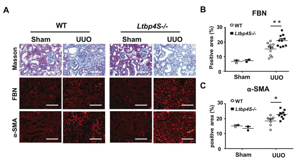
Fig. 2 Ltbp4S deficiency aggravates tubulointerstitial fibrosis in mice. Wild-type (WT) and Ltbp4S knockout (Ltbp4S−/−) mice were subjected to unilateral ureteral obstruction (UUO) to induce tubulointerstitial fibrosis for five days. A Representative histological images with Masson’s trichrome and immunofluorescence staining of kidneys after UUO. Scale bars, 100 μm. Computer-assisted quantitative analyses of histological images for fibronectin (B) and α-SMA (C) are shown. Data are presented as the mean ± SEM. n = 8–10 mice per group.
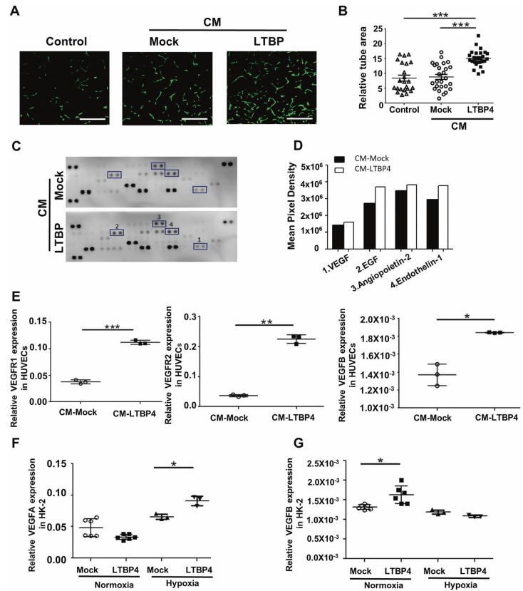
Fig. 4 LTBP4 facilitates angiogenesis in HUVECS and HK-2 cells. Conditioned media (CM) collected from Mock (CMMock) or LTBP4- overexpressing HEK293T cells (CM-LTBP4) were used to treat human umbilical vein endothelial cells (HUVECs) and human proximal tubule HK- 2 cells for 48 h, and tube areas and angiogenic factors were studied. A Representative images of tube formation in HUVECs. Control: HUVECs were treated with a regular growth medium. Scale bars, 400 μm. The results are from three independent experiments. B Densitometric analyses of tube areas in three groups are shown. C, D In proteomic profiling analysis, dot blot profiling for 55 angiogenesisrelated proteins showed relatively increasing expression of proangiogenic factors, including VEGF, EGF, angiopoietin-2 and endothelin-1. E Relative expression of VEGFRs and VEGF in HUVECs treated with CMMock and CM-LTBP4. CM-LTBP4 stimulated VEGFRs and VEGFB expression. F, G Effect of overexpression of LTBP4 in HK-2 cells. In normoxia, LTBP4 promoted VEGFB expression while, in hypoxia, LTBP4 enhanced VEGFA expression.
对野生型(WT)和Ltbp4S 基因敲除(ltbp 4s/)小鼠进行单侧输尿管梗阻(UUO ),诱导肾小管间质纤维化;与野生型小鼠相比,Ltbp4 缺陷导致更严重的肾小管间质纤维化TIF(图2A–C),提示LTBP4在肾纤维化过程中具有保护作用;
用从过度表达Mock 或LTBP4 的HEK293T 细胞收集的条件培养基处理HUVECs,发现细胞培养基中额外的LTBP4 显著促进HUVECs 中的管形成(图4A-C);为了阐明HUVECs 中受影响的血管生成分子,作者比较了用和不用条件培养基处理的细胞中血管生成相关标记物的表达。蛋白质组学分析显示,额外的LTBP4 处理刺激了HUVECs 中几种血管生成相关因子,包括血管内皮生长因子(VEGF)、表皮生长因子(EGF)、血管生成素-2 和内皮素-1(图4D);为了了解LTBP4 对血管生成信号的分子影响,作者研究了用额外LTBP4 处理的HUVECs 以及常氧和低氧LTBP4 过表达的HK-2 细胞中VEGFs和血管内皮生长受体(VEGFR)的表达。在含有LTBP4 的条件培养基存在下,HUVECs 中VEGFB 和VEGFR(包括VEGFR1 和VEGFR2 的表达上调(图4E);此外,发现LTBP4 刺激低氧HK-2 细胞中的VEGFA 表达(图4F),但VEGFB 表达未受到显著影响(图4G)。揭示LTBP4 诱导人脐静脉内皮细胞血管生成。